
Procedures We Perform
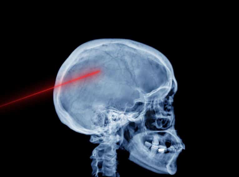
Craniotomy
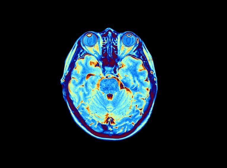
Craniotomy is a surgical procedure in which part of the skull is removed in order to view the brain. The piece of skull removed is called a “bone flap.” After the necessary brain surgery is performed, the bone flap is fitted back into the skull. Craniotomies are designated in different ways. A frontotemporal, parietal, temporal or suboccipital craniotomy is named after the bone that is removed. The minimally invasive “keyhole” craniotomy, on the other hand, is named after the small dime-sized incision that is made in the skull.
Reasons for Craniotomy
Craniotomies, which are performed by neurosurgeons, are appropriate in a number of situations, including for:
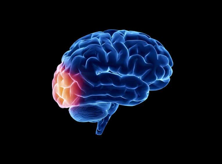
- Diagnosing/removing/treating brain tumors, including meningiomas
- Removing blood or blood clots, including subdural hematomas
- Repairing tears in the membrane that lines the brain
- Repairing or clipping aneurysms
- Draining brain abscesses
- Removing arteriovenous malformations (AVMs)
- Repairing skull fractures
- Repairing tears in the membrane lining the brain
- Treating epilepsy
- Implanting stimulator devices to treat movement disorders
Craniotomies are also used to relieve pressure within the brain caused by traumatic injury or stroke.
Craniotomy Procedure
Craniotomy is performed, sometimes under general anesthesia, in a hospital. An incision is made in the scalp (which is shaved at the incision site), and the bone flap is removed to allow access to the treatment area. There are cases in which patients remain awake during surgery, and are asked to move their legs, recite the alphabet or tell stories to ascertain whether brain functioning has been affected. The location of the incision will depend upon the area being treated. A medical drill may be used to create small holes (as in keyhole craniotomy), or a special saw may be used to cut the bone flap. Once the procedure is complete, any tissue that has been cut into is stitched together, and the bone flap is reattached using plates, sutures or wires.
Risk of Craniotomy
Surgery to treat a brain tumor is complex, and not without risks and complications. Some risks are related to the specific location being operated on. If, for example, the area of the brain being operated on controls speech, then speech may be affected. Aside from the usual surgical risks of infection and bleeding, other risks specific to craniotomy include swelling and accumulation of fluid, which can lead to brain damage and other serious complications. Surgery can also damage healthy brain tissue, causing problems with thinking, seeing and speaking.
Recovery from Craniotomy
Post-craniotomy, a patient usually has a headache for a few days, and can feel tired or weak. Most patients spend between 3 and 7 days in the hospital, but specific recovery times vary. Pain medication is prescribed in order to relieve symptoms and promote healing.
If malignant cells are found in the brain during the craniotomy, additional treatment may be necessary. Treatment may include radiation therapy, which uses high energy X-rays or gamma rays to destroy malignant cells, or chemotherapy, which uses drugs administered intravenously.
Additional Resources
Welcome to the office of neurosurgeon Dr. Vikas Rao, where your health comes first. Below are some of the neurosurgical treatments that we offer in Mission Viejo, CA:

Contact us today
Your concerns are important to us, and we want to make sure all of your questions are answered so you understand your options. Please contact our office with any questions, and our team will be happy to assist you.
Give us a call
We're open to serve
Book an appointment
Our doctor and staff are devoted to our patients. Please fill out the form below with any questions or to schedule an appointment and our team will get back to you within 24 to 48 hours.